


New brain atlas gives researchers a closer look at the machinery of movement
A collection of 27 papers maps cells in the basal ganglia, building a shared reference for studying development and neurological conditions.
How this story was made
This story was written with AI assistance from National Institutes of Health’s source documents. Its figures and quotations were checked against their sources, and Digitalage Desk reviewed and approved it before publication. It is published under the name of Peter Michaels, Editor.
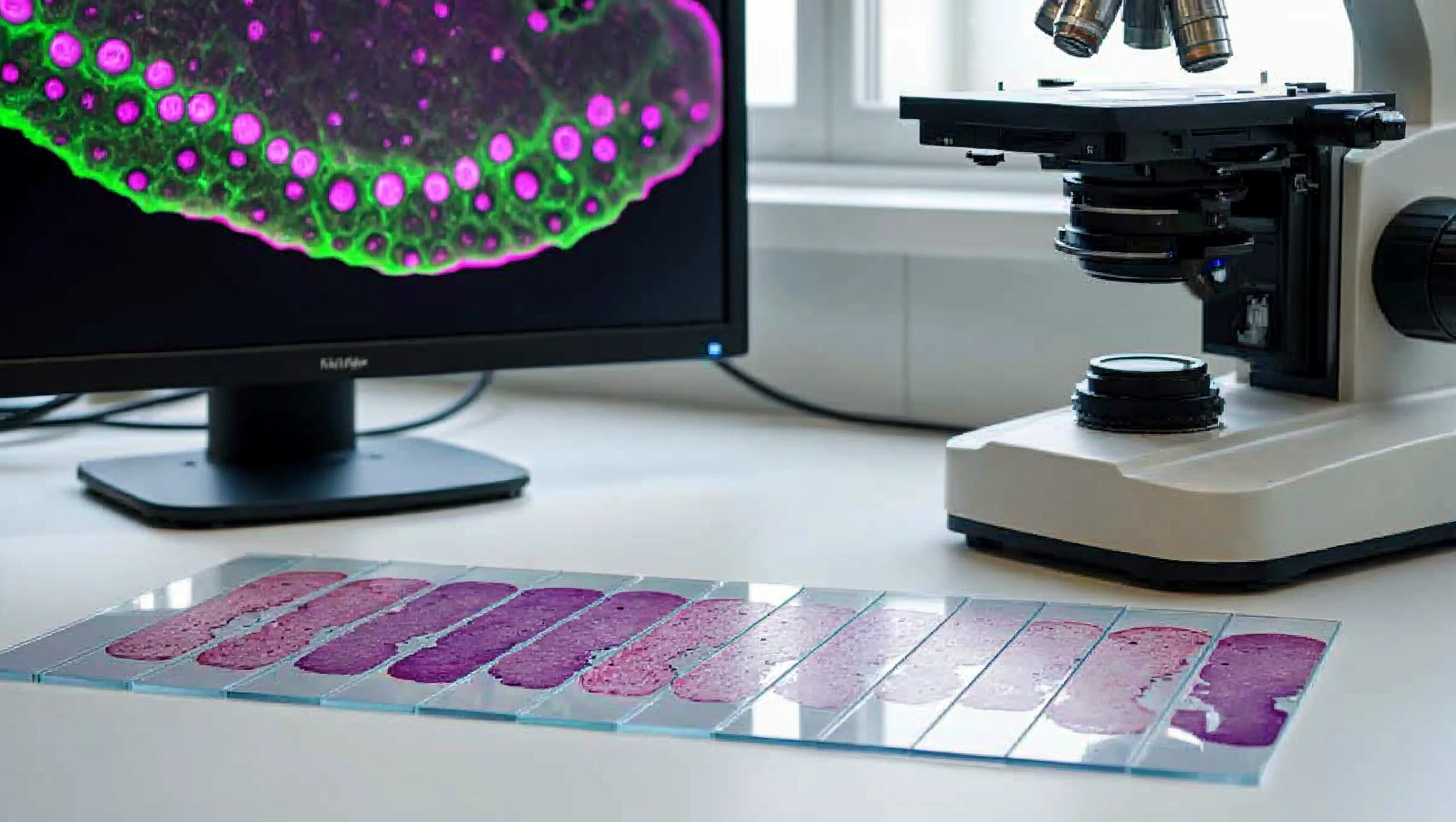
Scientists have produced a detailed cellular atlas of the basal ganglia, the brain structures involved in deliberate movement, learning and focus, giving researchers a new way to examine how those systems develop and change in disease.
The National Institutes of Health announced the work on November 12, describing a collection of 27 papers from the BRAIN Initiative Cell Atlas Network. Instead of treating a brain region as a single block, the project distinguishes its component cell populations and places them in three-dimensional space.
Researchers combined single-cell sequencing of donated postmortem human tissue with advanced imaging. The resulting maps cover neurons and supporting glial cells, including developmental stages, with related atlases from other primates and mice.
Primary sourceNational Institutes of Health: “For the first time, researchers map out the brain's behavioral gatekeeper”According to the network’s program description, its wider objective is to create reference maps across species and the lifespan. The program builds on an earlier cell census, extending the work from identifying cell types toward locating them and understanding their interactions. Its resources are intended for use throughout the research community.
That common framework matters when laboratories compare findings from human tissue with experiments in animals. It can help investigators identify a corresponding cell population rather than assume that an entire region behaves identically across species. It does not eliminate differences between an animal model and a human condition.
Data processing is part of making those comparisons useful. The network helped develop Optimus, an open-source analysis workflow that converts sequencing reads into cell-by-cell gene counts and quality measures. Its documentation describes support for human and mouse data, allowing other researchers to inspect and reuse the computational approach.
According to NIH, one study used the reference to organize postmortem sequencing data from people with autism, identifying differences in cell populations within the striatum. That is a research finding about tissue, not a diagnostic test for an individual.
The immediate result is a set of maps and tools other scientists can use. Establishing whether particular cellular differences cause symptoms, and whether changing them helps patients, requires further experiments beyond constructing the atlas.
End of story